File:Seminal vesicle high mag.jpg

Contents
Summary
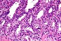
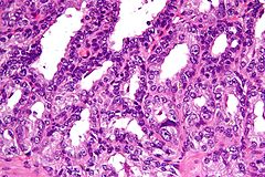
High magnification <a href="https://en.wikipedia.org/wiki/micrograph" class="extiw" title="w:micrograph">micrograph</a> of <a href="https://en.wikipedia.org/wiki/seminal_vesicle" class="extiw" title="w:seminal vesicle">seminal vesicle</a>. <a href="https://en.wikipedia.org/wiki/H%26E_stain" class="extiw" title="w:H&E stain">H&E stain</a>. <a href="https://en.wikipedia.org/wiki/prostatectomy" class="extiw" title="w:prostatectomy">Prostatectomy specimen</a>.
The epithelium of seminal vesicle is the same as <a href="https://en.wikipedia.org/wiki/ejaculatory_duct" class="extiw" title="w:ejaculatory duct">ejaculatory duct</a>.<a href="#cite_note-pmid12657938-1">[1]</a>
References
- <a href="#cite_ref-pmid12657938_1-0">↑</a> Leroy X, Ballereau C, Villers A, et al. (April 2003). "MUC6 is a marker of seminal vesicle-ejaculatory duct epithelium and is useful for the differential diagnosis with prostate adenocarcinoma". Am. J. Surg. Pathol. 27 (4): 519–21. <a href="https://en.wikipedia.org/wiki/PMID" class="extiw" title="w:PMID">PMID</a> <a rel="nofollow" class="external text" href="http://www.ncbi.nlm.nih.gov/pubmed/12657938">12657938</a>.
Related images
-
<a href="//commons.wikimedia.org/wiki/File:Seminal_vesicle_low_mag.jpg" class="image"><img alt="" src="https://upload.wikimedia.org/wikipedia/commons/thumb/8/89/Seminal_vesicle_low_mag.jpg/80px-Seminal_vesicle_low_mag.jpg" width="80" height="120" srcset="https://upload.wikimedia.org/wikipedia/commons/thumb/8/89/Seminal_vesicle_low_mag.jpg/120px-Seminal_vesicle_low_mag.jpg 1.5x, https://upload.wikimedia.org/wikipedia/commons/thumb/8/89/Seminal_vesicle_low_mag.jpg/160px-Seminal_vesicle_low_mag.jpg 2x" data-file-width="2848" data-file-height="4272"></a>
Low mag.
-
<a href="//commons.wikimedia.org/wiki/File:Seminal_vesicle_intermed_mag.jpg" class="image"><img alt="" src="https://upload.wikimedia.org/wikipedia/commons/thumb/7/7c/Seminal_vesicle_intermed_mag.jpg/120px-Seminal_vesicle_intermed_mag.jpg" width="120" height="80" srcset="https://upload.wikimedia.org/wikipedia/commons/thumb/7/7c/Seminal_vesicle_intermed_mag.jpg/180px-Seminal_vesicle_intermed_mag.jpg 1.5x, https://upload.wikimedia.org/wikipedia/commons/thumb/7/7c/Seminal_vesicle_intermed_mag.jpg/240px-Seminal_vesicle_intermed_mag.jpg 2x" data-file-width="4272" data-file-height="2848"></a>
Intermed. mag.
-
<a href="//commons.wikimedia.org/wiki/File:Seminal_vesicle_high_mag.jpg" class="image"><img alt="" src="https://upload.wikimedia.org/wikipedia/commons/thumb/2/2b/Seminal_vesicle_high_mag.jpg/120px-Seminal_vesicle_high_mag.jpg" width="120" height="80" srcset="https://upload.wikimedia.org/wikipedia/commons/thumb/2/2b/Seminal_vesicle_high_mag.jpg/180px-Seminal_vesicle_high_mag.jpg 1.5x, https://upload.wikimedia.org/wikipedia/commons/thumb/2/2b/Seminal_vesicle_high_mag.jpg/240px-Seminal_vesicle_high_mag.jpg 2x" data-file-width="4272" data-file-height="2848"></a>
High mag.
Licensing
Lua error in package.lua at line 80: module 'strict' not found.
File history
Click on a date/time to view the file as it appeared at that time.
| Date/Time | Thumbnail | Dimensions | User | Comment | |
|---|---|---|---|---|---|
| current | 12:08, 7 January 2017 | | 4,272 × 2,848 (5.42 MB) | 127.0.0.1 (talk) | High magnification <a href="https://en.wikipedia.org/wiki/micrograph" class="extiw" title="w:micrograph">micrograph</a> of <b><a href="https://en.wikipedia.org/wiki/seminal_vesicle" class="extiw" title="w:seminal vesicle">seminal vesicle</a></b>. <a href="https://en.wikipedia.org/wiki/H%26E_stain" class="extiw" title="w:H&E stain">H&E stain</a>. <a href="https://en.wikipedia.org/wiki/prostatectomy" class="extiw" title="w:prostatectomy">Prostatectomy specimen</a>. <p>The epithelium of seminal vesicle is the same as <a href="https://en.wikipedia.org/wiki/ejaculatory_duct" class="extiw" title="w:ejaculatory duct">ejaculatory duct</a>.<sup id="cite_ref-pmid12657938_1-0" class="reference"><a href="#cite_note-pmid12657938-1">[1]</a></sup></p> <h2><span class="mw-headline" id="References">References</span></h2> <div class="reflist references-column-count references-column-count-1" style="-moz-column-count: 1; -webkit-column-count: 1; column-count: 1; list-style-type: decimal;"> <ol class="references"> <li id="cite_note-pmid12657938-1"> <span class="mw-cite-backlink"><a href="#cite_ref-pmid12657938_1-0">↑</a></span> <span class="reference-text"><cite style="font-style:normal">Leroy X, Ballereau C, Villers A, <i>et al.</i> (April 2003). "MUC6 is a marker of seminal vesicle-ejaculatory duct epithelium and is useful for the differential diagnosis with prostate adenocarcinoma". <i>Am. J. Surg. Pathol.</i> <b>27</b> (4): 519–21. <a href="https://en.wikipedia.org/wiki/PMID" class="extiw" title="w:PMID">PMID</a> <a rel="nofollow" class="external text" href="http://www.ncbi.nlm.nih.gov/pubmed/12657938">12657938</a>.</cite></span> </li> </ol> </div> <h2><span class="mw-headline" id="Related_images">Related images</span></h2> <ul class="gallery mw-gallery-traditional"> <li class="gallerybox" style="width: 155px"><div style="width: 155px"> <div class="thumb" style="width: 150px;"><div style="margin:15px auto;"><a href="//commons.wikimedia.org/wiki/File:Seminal_vesicle_low_mag.jpg" class="image"><img alt="" src="https://upload.wikimedia.org/wikipedia/commons/thumb/8/89/Seminal_vesicle_low_mag.jpg/80px-Seminal_vesicle_low_mag.jpg" width="80" height="120" srcset="https://upload.wikimedia.org/wikipedia/commons/thumb/8/89/Seminal_vesicle_low_mag.jpg/120px-Seminal_vesicle_low_mag.jpg 1.5x, https://upload.wikimedia.org/wikipedia/commons/thumb/8/89/Seminal_vesicle_low_mag.jpg/160px-Seminal_vesicle_low_mag.jpg 2x" data-file-width="2848" data-file-height="4272"></a></div></div> <div class="gallerytext"> <p>Low mag. </p> </div> </div></li> <li class="gallerybox" style="width: 155px"><div style="width: 155px"> <div class="thumb" style="width: 150px;"><div style="margin:35px auto;"><a href="//commons.wikimedia.org/wiki/File:Seminal_vesicle_intermed_mag.jpg" class="image"><img alt="" src="https://upload.wikimedia.org/wikipedia/commons/thumb/7/7c/Seminal_vesicle_intermed_mag.jpg/120px-Seminal_vesicle_intermed_mag.jpg" width="120" height="80" srcset="https://upload.wikimedia.org/wikipedia/commons/thumb/7/7c/Seminal_vesicle_intermed_mag.jpg/180px-Seminal_vesicle_intermed_mag.jpg 1.5x, https://upload.wikimedia.org/wikipedia/commons/thumb/7/7c/Seminal_vesicle_intermed_mag.jpg/240px-Seminal_vesicle_intermed_mag.jpg 2x" data-file-width="4272" data-file-height="2848"></a></div></div> <div class="gallerytext"> <p>Intermed. mag. </p> </div> </div></li> <li class="gallerybox" style="width: 155px"><div style="width: 155px"> <div class="thumb" style="width: 150px;"><div style="margin:35px auto;"><a href="//commons.wikimedia.org/wiki/File:Seminal_vesicle_high_mag.jpg" class="image"><img alt="" src="https://upload.wikimedia.org/wikipedia/commons/thumb/2/2b/Seminal_vesicle_high_mag.jpg/120px-Seminal_vesicle_high_mag.jpg" width="120" height="80" srcset="https://upload.wikimedia.org/wikipedia/commons/thumb/2/2b/Seminal_vesicle_high_mag.jpg/180px-Seminal_vesicle_high_mag.jpg 1.5x, https://upload.wikimedia.org/wikipedia/commons/thumb/2/2b/Seminal_vesicle_high_mag.jpg/240px-Seminal_vesicle_high_mag.jpg 2x" data-file-width="4272" data-file-height="2848"></a></div></div> <div class="gallerytext"> <p>High mag. </p> </div> </div></li> </ul> |
- You cannot overwrite this file.
File usage
The following page links to this file: