Fusiform gyrus
| Fusiform gyrus | |
|---|---|

Medial surface of left cerebral hemisphere. (Fusiform gyrus shown in orange)
|
|

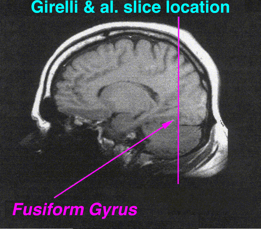
Medial surface of right cerebral hemisphere. (Fusiform gyrus visible near bottom)
|
|
| Details | |
| Latin | gyrus fusiformis |
| Identifiers | |
| NeuroNames | hier-121 |
| NeuroLex ID | Fusiform Gyrus |
| Dorlands /Elsevier |
g_13/12405287 |
| TA | Lua error in Module:Wikidata at line 744: attempt to index field 'wikibase' (a nil value). |
| TH | {{#property:P1694}} |
| TE | {{#property:P1693}} |
| FMA | {{#property:P1402}} |
| Anatomical terms of neuroanatomy
[[[d:Lua error in Module:Wikidata at line 863: attempt to index field 'wikibase' (a nil value).|edit on Wikidata]]]
|
|
The fusiform gyrus, also known as the (discontinuous) occipitotemporal gyrus, is part of the temporal lobe and occipital lobe in Brodmann area 37.[1] The fusiform gyrus is located between the lingual gyrus and parahippocampal gyrus above, and the inferior temporal gyrus below.[2] Though the functionality of the fusiform gyrus is not fully understood, it has been linked with various neural pathways related to recognition. Additionally, it has been linked to various neurological phenomena such as synesthesia, dyslexia, and prosopagnosia.
Contents
Anatomy
The lateral and medial portions are separated by the shallow mid-fusiform sulcus.[3][4][5]
History
The fusiform gyrus has a contentious history that has recently been clarifed.[6] Emil Huschke first labeled the fusiform gyrus in 1854. Huschke also labeled the lingual gyrus, as well as the lingual sulcus. Burt Wilder disagreed with the fusiform label because it was too descriptive in nature. Instead, he wanted to change the name to the subcollateral gyrus because the fusiform gyrus is inferior to the collateral sulcus. Wilhelm His, Sr. disagreed with Wilder and sided with Huschke. The fusiform label still persists today.
Function
The exact functionality of the fusiform gyrus is still disputed, but there is relative consensus on its involvement in the following pathways:
Processing of color information
(see Color center)
In 2003, V.S. Ramachandran collaborated with scientists from the Salk Institute for Biological Studies in order to identify the potential role of the fusiform gyrus within the color processing pathway in the brain. Examining the relationship within the pathway specifically in cases of synesthesia, Ramachandran found that synesthetes on average have a higher density of fibers surrounding the angular gyrus. The angular gyrus is involved in higher processing of colors.[7] The fibers relay shape information from the fusiform gyrus to the angular gyrus in order to produce the association of colors and shapes in grapheme-color synesthesia.[7] Cross-activation between the angular and fusiform gyri has been observed in the average brain, implying that the fusiform gyrus regularly communicates with the visual pathway.[8]
Face and body recognition
(see Fusiform face area)
Brain imaging techniques have identified activity in this region while subjects observe faces. This has led to subsequent research studying the effect of stimulating the region with electrodes.[9] However, many scientists counter that this observation is only a correlation and that it does not indicate that the fusiform gyrus is the sole region involved in facial recognition. Additionally, the fusiform gyrus has been seen to have influence on the amygdala response to emotional faces.[10]
Word recognition
(see Visual word form area)
It is believed that the neurons in the left portion of the fusiform gyrus are used in word recognition. Similar to grapheme-color synesthesia, many synesthetes perceive certain words as possessing a distinct color or hue.[11]
Within-category identification
After further research by scientists at MIT, it was concluded that both the left and right fusiform gyrus played different roles from one another, but were subsequently interlinked. The left fusiform gyrus plays the role of recognizing "face-like" features in objects that may or may not be actual faces whereas the right fusiform gyrus plays the role in determining whether or not the recognized "face-like" feature is, in fact, an actual face.[12]
Associated neurological phenomena
The fusiform gyrus has been speculated to be associated with various neurological phenomena. Many are outlined below:
Prosopagnosia
Some researchers think that the fusiform gyrus may be related to the disorder known as prosopagnosia, or face blindness. Research has also shown that the fusiform face area, the area within the fusiform gyrus, is heavily involved in face perception but only to any generic within-category identification that is shown to be one of the functions of the fusiform gyrus.[13] Abnormalities of the fusiform gyrus have also been linked to Williams syndrome.[14] Fusiform gyrus has also been involved in the perception of emotions in facial stimuli.[15] However, individuals with autism show little to no activation in the fusiform gyrus in response to seeing a human face.[16]
Synaesthesia
Recent research has seen activation of the fusiform gyrus during subjective grapheme-color perception in people with synaesthesia.[17] The effect of the fusiform gyrus in grapheme sense seems somewhat more clear as the fusiform gyrus seems to play a key role in word recognition. The connection to color may be due to cross wiring of (being directly connected to) areas of the fusiform gyrus and other areas of the visual cortex associated with experiencing color.[10]
Dyslexia
For those with dyslexia, it has been seen that the fusiform gyrus is underactivated and has reduced gray matter density.[18]
Face hallucinations
Increased neurophysiological activity in the fusiform face area may produce hallucinations of faces, whether realistic or cartoonesque, as seen in Charles Bonnet syndrome, hypnagogic hallucinations, peduncular hallucinations, or drug-induced hallucinations.[19]
References
- ↑ Nature Neuroscience, vol7, 2004
- ↑ Lua error in package.lua at line 80: module 'strict' not found.
- ↑ Lua error in package.lua at line 80: module 'strict' not found.
- ↑ Lua error in package.lua at line 80: module 'strict' not found.
- ↑ Lua error in package.lua at line 80: module 'strict' not found.
- ↑ Lua error in package.lua at line 80: module 'strict' not found.
- ↑ 7.0 7.1 Lua error in package.lua at line 80: module 'strict' not found.
- ↑ Lua error in package.lua at line 80: module 'strict' not found.
- ↑ Lua error in package.lua at line 80: module 'strict' not found.
- ↑ 10.0 10.1 Lua error in package.lua at line 80: module 'strict' not found.
- ↑ Lua error in package.lua at line 80: module 'strict' not found.
- ↑ Trafton, A. "How does our brain know what is a face and what’s not?" MIT News
- ↑ McCarthy, G et al. Face-specific processing in the fuman fusform gyrus.J. Cognitive Neuroscicence. 9, 605–610(1997).
- ↑ A. L. Reiss, et al. Preliminary Evidence Of Abnormal White Matter Related To The Fusiform Gyrus In Williams Syndrome: A Diffusion Tensor Imaging Tractography Study.Genes, Brain & Behavior 11.1, 62–68(2012)
- ↑ Lua error in package.lua at line 80: module 'strict' not found.
- ↑ Lua error in package.lua at line 80: module 'strict' not found.
- ↑ Imaging of connectivity in the synaesthetic brain « Neurophilosophy
- ↑ Lua error in package.lua at line 80: module 'strict' not found.
- ↑ Jan Dirk Blom. A Dictionary of Hallucinations. Springer, 2010, p. 187. ISBN 978-1-4419-1222-0
Additional images
-

Fusiform gyrus
-

Fusiform gyrus animation
-

Cerebrum.Inferior view. Deep dissection
-

Fusiform gyrus in a ventral view (from below, diagrammatic), labeled at left
-

Fusiform gyrus seen in a ventral view


External links
| Wikimedia Commons has media related to Fusiform gyrus. |
- Atlas image: n1a2p13 at the University of Michigan Health System – "Cerebral Hemisphere, Inferior View"
- Location at mattababy.org
- "VS Ramachandran on your mind" at ted.com
- "Oliver Sacks: What hallucination reveals about our minds" at ted.com
- NIF Search – Fusiform Gyrus via the Neuroscience Information Framework