Esophageal gland
From Infogalactic: the planetary knowledge core
| Esophageal glands | |
|---|---|
| File:Illu esophageal layers.jpg
Layers of Esophageal Wall:
1. Mucosa 2. Submucosa 3. Muscularis 4. Adventitia 5. Striated muscle 6. Striated and smooth 7. Smooth muscle 8. Lamina muscularis mucosae 9. Esophageal glands |
|

Section of the human esophagus. Moderately magnified. The section is transverse and from near the middle of the gullet.
a. Fibrous covering. b. Divided fibers of longitudinal muscular coat. c. Transverse muscular fibers. d. Submucous or areolar layer. e. Muscularis mucosae. f. Mucous membrane, with vessels and part of a lymphoid nodule. g. Stratified epithelial lining. h. Mucous gland. i. Gland duct. m’. Striated muscular fibers cut across. |
|
| Details | |
| Latin | glandulae oesophageae |
| Identifiers | |
| Dorlands /Elsevier |
g_06/12392509 |
| TA | Lua error in Module:Wikidata at line 744: attempt to index field 'wikibase' (a nil value). |
| TH | {{#property:P1694}} |
| TE | {{#property:P1693}} |
| FMA | {{#property:P1402}} |
| Anatomical terminology
[[[d:Lua error in Module:Wikidata at line 863: attempt to index field 'wikibase' (a nil value).|edit on Wikidata]]]
|
|
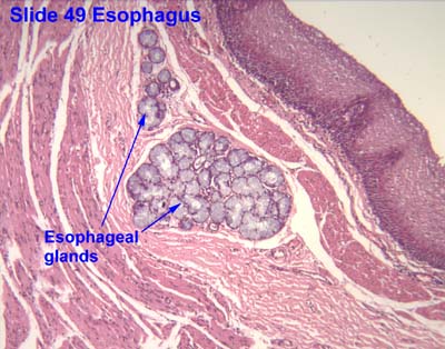
The esophageal glands are small compound racemose exocrine glands of the mucous type.
There are two types:
- Esophageal glands proper- located in the submucosa and secrete acid mucin for lubrication
- Esophageal cardiac glands- in the lamina propria and secrete neutral mucus that protects the esophagus from acidic gastric juices.
Each opens upon the surface by a long excretory duct.
References
This article incorporates text in the public domain from the 20th edition of Gray's Anatomy (1918)
External links
- Histology image: 49_07 at the University of Oklahoma Health Sciences Center
- Histology image: 10802loa – Histology Learning System at Boston University
<templatestyles src="Asbox/styles.css"></templatestyles>
|
This human digestive system article is a stub. You can help Wikipedia by expanding it. |